Choosing the Right Method for Spider Vein Treatment

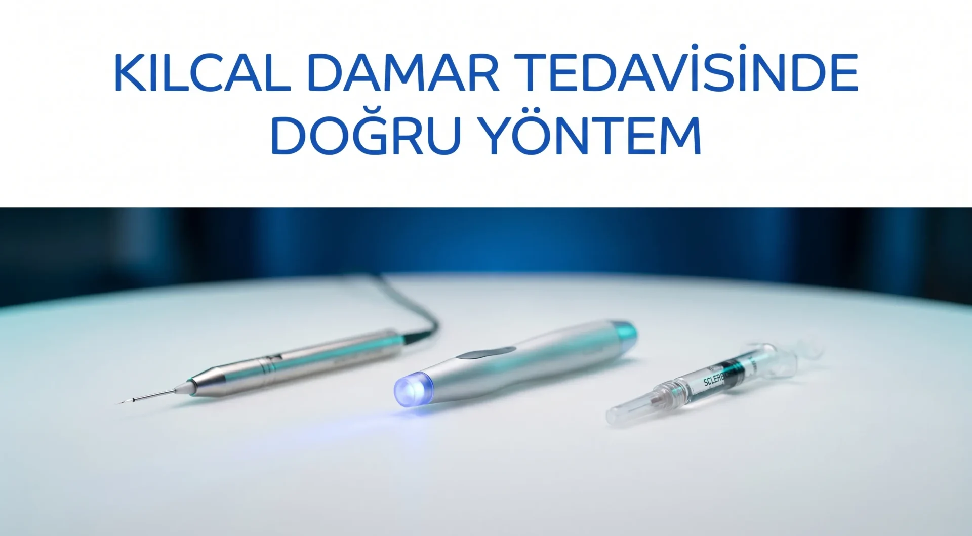
There is no single universal correct method in spider vein treatment; the right one is simply the most appropriate for that patient, that vessel, and that skin.
In this post, I want to share what determines method selection in capillary vein treatment, common mistakes I see, and how I make this decision in my practice.
Why Is There No Single Correct Method?
Capillary veins, or telangiectasias, may look similar on the surface, but they differ enormously in vessel diameter, depth, the presence of feeder veins, and the structure of the surrounding skin. A superficial spider vein 0.1 mm in diameter and a red network fed by a rooted reticular vein 2-3 mm in diameter will not produce the same result with the same treatment.1
Moreover, a method that works in one patient may produce inadequate results or unwanted effects in another with different skin tone or vascular anatomy. For this reason, treatment selection starts with a physical examination, continues with Doppler ultrasound, and is only finalised after both.
Clinical note: With every patient who tells me “I had capillary vein treatment but it didn’t work,” my first question is: “Was a Doppler scan done beforehand?” The answer is usually no. Yet if there is an undetected feeder vessel behind the superficial capillaries, treating the surface without treating the source is like painting the wall while the floor is leaking underneath.
Method Selection Criteria
When deciding which method to recommend for a patient, I assess the following criteria:
| Assessment Criterion | Clinical Significance |
|---|---|
| Vessel diameter | 0.1-0.5 mm: radiofrequency or laser; 0.5-3 mm: sclerotherapy is the primary option |
| Vessel depth | Superficial: heat-based methods; deep: injectable methods are more effective |
| Skin tone (Fitzpatrick) | Darker skin (type IV-VI): higher pigmentation risk, energy setting is critical |
| Presence of feeder vessel | If reticular vein feeding is present, the source must be treated first |
| Location | Face (especially around the nose): laser; legs: sclerotherapy or RF |
| Previous treatment history | Recurrent cases may require a change in method |
| Patient’s general health | Contraindications such as coagulation disorders, sclerosant allergy |
All of these assessments, both clinical and ultrasonographic findings, form the foundation of the method decision. For more detailed information, you can visit my capillary vein treatment page.
I sometimes receive this question: “Doctor, could I just choose based on price after reading online?” Cost is certainly a factor, but it should not be the deciding one. A cheap procedure done with the wrong method can turn into paying twice the second time around with the right one. Furthermore, if treatment-related pigmentation or burns occur, correcting these requires far longer and more costly processes.
Commonly Used Methods
Radiofrequency Thermocoagulation
One of the methods that has come to the fore in recent years is radiofrequency thermocoagulation. A fine electrode tip is brought into contact with or very close to the vessel; controlled thermal energy coagulates the vessel wall and the vessel closes. It does not require injection, which makes it preferable in patients with injection anxiety or for very fine vessels (0.1-0.3 mm).
However, the strength of this method is also its greatest trap: if too much energy is delivered, surrounding tissue is damaged; if too little, the vessel is not sufficiently coagulated. Achieving the right result requires a precise energy protocol calibrated to the patient’s skin type.
For a broader look at this topic, you can read my post on the advantages of radiofrequency treatment for spider veins.
Laser Treatment
Laser is an effective option particularly for fine vessels and redness in the facial area. Light energy is selectively absorbed by haemoglobin within the vessel and converted to heat. The main limitation: in darker skin tones (Fitzpatrick type IV and above), the risk of pigmentation increases; these patients require lower energy settings and cooling systems.
Sclerotherapy
One of the most widely used methods for capillary and spider veins on the legs is sclerotherapy. A chemical agent (such as polidocanol or sodium tetradecyl sulphate) is injected into the vessel through a fine needle; the vessel wall becomes irritated and closes.2 For vessels over 0.5 mm, particularly in networks fed by reticular veins, sclerotherapy generally covers a larger area in a single session compared with radiofrequency.
For comprehensive information on sclerotherapy, you can visit my capillary treatment page.
The Importance of Patient Selection
No matter how good the method, when applied to an unsuitable patient it produces disappointing results. Points that must not be overlooked in patient selection:
- Status of the feeder vessel: If the capillary veins are fed by an invisible but incompetent reticular vein, superficial treatments performed without treating the source yield temporary results. Doppler ultrasound assessment is therefore essential.
- Active skin diseases: Treatment is not applied to areas of eczema, dermatitis, or infection.
- Coagulation disorders: In patients using blood thinners or with coagulation disorders, risk assessment is required before sclerotherapy.
- Pregnancy and breastfeeding: Elective cosmetic procedures are deferred during this period.
- Realistic expectations: Capillary vein treatment closes existing vessels; however, it cannot permanently prevent new vessel formation. If genetic predisposition, hormonal changes, or lifestyle factors come into play, new vessels may form.3
Why Is Correct Energy Setting Critical?
The amount of energy used in radiofrequency and laser treatments directly determines both efficacy and safety. Excessive energy leads to:
- Thermal damage to surrounding tissue
- Skin surface burns
- Dark or light pigmentation changes (hyperpigmentation or hypopigmentation)
Insufficient energy, on the other hand, does not close the vessel; either the same vessel reopens within a short time or it appears to resolve during the first few weeks and then recurs.
The energy protocol is determined according to skin type (Fitzpatrick scale), vessel diameter, location, and the technical specifications of the device used. There is therefore no “standard” setting; every session is individual to the patient.
Even in the same patient, different areas may require different energy levels: a fine vessel on the inner surface of the leg and a deeper vessel below the knee are not treated with the same settings. An experienced practitioner assesses and manages this difference in real time during the session. Because there is no standard template, experience in this field becomes a critical factor determining both efficacy and safety.
Adverse Effects Seen with Inadequate Application
Problems that may be encountered with the wrong method, wrong patient selection, or incorrect energy application include:
- Telangiectatic matting: Expansion of the capillary network in the area where the vessel has closed following treatment. Seen particularly with excessively concentrated sclerosant or high-energy applications.4
- Hyperpigmentation: Dark discolouration along the track of the closed vessel. Generally resolves within 6-12 months; however, it may be more persistent in darker skin tones.
- Skin burns and crusting: Seen particularly with high energy use in radiofrequency or laser procedures.
- Recurrence and “rebound”: In cases where the feeder vessel has not been closed, the capillary network quickly refills. This is the result of inadequate assessment, not of the method itself.
For this reason, assessing the outcome after treatment is at least as important as the treatment itself. In unmonitored cases, neither the patient’s expectations are met nor is a decision to change method made in time when necessary.
The following timeline is important for long-term outcome evaluation: redness and mild swelling immediately after treatment are normal and generally resolve within a few days. Complete disappearance of the treated vessel may take 4-8 weeks. For this reason, it is necessary to wait at least 6-8 weeks before declaring a treatment “unsuccessful.” Rushing to evaluate results can be misleading; both early satisfaction and early disappointment often stem from not observing this period.
Post-treatment daily life restrictions for the patient also vary by method. After sclerotherapy, compression stockings and brief walking restrictions are usually recommended; after radiofrequency, protecting the area from sunlight is important. Adhering to these recommendations both accelerates healing and reduces the risk of adverse effects.
Frequently Asked Questions
How many sessions are needed for capillary vein treatment?
This depends entirely on the extent of the vessels, the chosen method, and the patient’s skin type. For fine vessels in a small area, 1-2 sessions may be sufficient, while more sessions may be required for large areas or cases requiring feeder vessel treatment. The number of sessions is not given as a fixed figure from the outset; the plan is updated based on the outcome of the first session.
Is radiofrequency or sclerotherapy better?
The two are not rivals but complementary methods aimed at different vessel types. For very fine (0.1-0.3 mm) and superficial vessels, radiofrequency stands out, while for leg vessels over 0.5 mm and spider vein networks, sclerotherapy generally offers the advantage of wider coverage. The choice is determined by clinical examination and ultrasound assessment.
Is treatment permanent?
The treated vessel closes permanently. However, if genetic predisposition, hormonal changes, or prolonged standing continue, new vessels may form at different locations. Treatment resolves the current problem; it does not guarantee prevention of new vessel formation.
Is treatment painful?
In radiofrequency thermocoagulation, there is a brief stinging and mild burning sensation. In sclerotherapy, mild pain during injection is normal. In both cases, pain is generally very brief; topical anaesthesia options are available.
Which skin tones require extra caution?
In patients with Fitzpatrick type IV-VI skin (olive to dark), the risk of hyperpigmentation with heat-based methods increases significantly. These patients require lower energy, more widely spaced applications, and the use of sun protection. This does not mean treatment cannot be performed; but the protocol must be carefully adapted.
For international patients: If you are considering varicose vein treatment in Turkey, our İzmir clinic offers comprehensive evaluation and tailored protocols. For spider vein treatment Turkey, visit our treatment overview for detailed information.
References
- Sundaram M, et al. Superficial Venous Disease – An Updated Review. Dermatol Surg. 2024. PubMed PMID: 38583765. https://pubmed.ncbi.nlm.nih.gov/38583765/
- Alós J, et al. Skin hyperpigmentation after sclerotherapy with polidocanol: A systematic review. Phlebology. 2022. PubMed PMID: 36196455. https://pubmed.ncbi.nlm.nih.gov/36196455/
- NHS. Varicose veins – Treatment. https://www.nhs.uk/conditions/varicose-veins/treatment/
- NICE. Ultrasound-guided foam sclerotherapy for varicose veins. Interventional Procedures Guidance IPG440. https://www.nice.org.uk/guidance/ipg440
- Elvis AM, Ekta JS. Ozone therapy: A clinical review. J Nat Sci Biol Med. 2011;2(1):66-70. PubMed PMID: 22470237. https://pubmed.ncbi.nlm.nih.gov/22470237/
Op. Dr. Onur Üstünel
Cardiovascular Surgery Specialist