
Are Spider Vein Treatments Permanent?

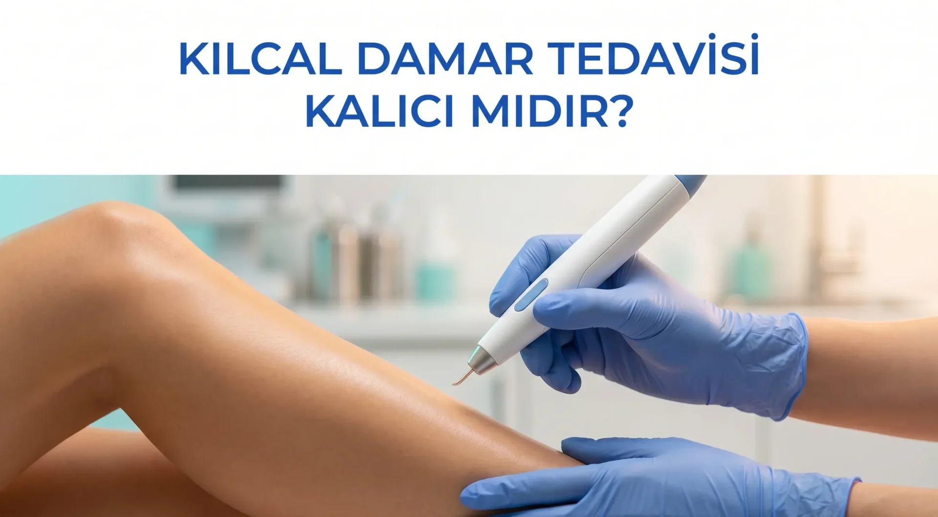
A patient has invested time and effort in treatment; what they want is to see a lasting result. My answer is always an honest one: for the treated vessels, generally yes, but when it comes to new vessel formation, the process requires monitoring.
In this article I want to share how spider vein treatment works, the factors that affect permanence, and what to pay attention to for long-term success.
Let me say this upfront: the question “Is it permanent?” actually contains two separate questions. The first is whether the treated vessels return. The second is whether new spider veins will form. The answer to the first question is generally positive; the second depends on an individual’s genetic makeup, risk factors, and the scope of treatment.
Do Treated Vessels Come Back?
In a successful spider vein treatment, the targeted vessels disappear permanently. The methods used in capillary vein treatment damage the wall of the target vessel, turning it into fibrotic tissue; the vessel loses its function and the body gradually resorbs it.1
In sclerotherapy, for example, the solution injected into the vessel damages the vascular endothelium to achieve closure. The radiofrequency method creates a similar effect with thermal energy. In both cases, the treated vessel is permanently rendered non-functional and does not return.2
So if a vessel reappears on the patient’s leg after treatment, it is most likely not the same vessel. It is a new vessel that has developed at a different point, or grown from a small branch that the treatment could not reach.
In sclerotherapy, the success rate depends on using the agent at the correct concentration and volume, the injection technique, and the size of the vessel being treated. Very thin capillaries may require a lower concentration, while wider branches connecting to the feeder vein require a different protocol. This nuance is decisive for long-term outcomes.2
Important distinction: When speaking about “permanence,” two things must be distinguished: the closure of the treated vessel, which is generally permanent; and the prevention of new spider vein formation, which varies from person to person and is a separate process that requires monitoring.
Why Do New Spider Veins Form?
The most honest answer to this question is: if the underlying cause has not been treated, new vessels may form.
Spider veins (telangiectasias in medical terminology) may appear superficial but carry an underlying source. This source is small feeder veins or a deeper valvular insufficiency. When we look at the difference between spider veins and varicose veins, this relationship becomes very clear: a significant proportion of spider veins are the superficial reflection of deeper venous insufficiency.
If the feeder vein is identified and not included in the treatment, new spider veins can form over time in the same area. This does not mean the treatment has failed; it means the underlying problem is continuing.1
I explain this to my patients as follows: closing spider veins one by one is like plucking the leaves of a tree one by one. As long as the tree continues to be nourished, new leaves will grow. Cutting off the tree’s nourishment, that is, closing the feeder vein, forms the foundation of long-term results. For this reason, in some patients it may be necessary to close a somewhat larger feeder vein before beginning spider vein treatment. This step is taken first, after which the spider veins respond much better to treatment.
Additionally, in some areas of the body, particularly behind the knee and on the inner side of the calf, the feeder venous system can form a more complex network. For this reason, some patients may require more than one session, and allowing adequate waiting time between all treatment sessions is also important.2
Skin Structure and Genetic Predisposition
Individual differences in spider vein formation are very pronounced. I can see completely different clinical pictures in two people with the same risk factors. This is largely due to genetic makeup.
In individuals with weak connective tissue in the skin and genetically thin vessel walls, spider veins appear earlier and there is a higher likelihood of new ones developing after treatment. This is not a failure, it is the management of a chronic tendency.
I should also add regarding connective tissue: a genetic predisposition toward connective tissue weakness can affect the vessel wall structure in many parts of the body. Such patients may respond well to spider vein treatment but see new formations in different areas. This is not a failure but the individual expression of a chronic process. Explaining this to the patient from the outset allows them to develop realistic expectations of treatment.
Some patients say “It improved but came back after a year or two.” The most common explanations for this situation are:
- Protective measures were not continued after treatment (compression stockings, avoiding prolonged standing, avoiding heat exposure)
- The underlying feeder vein or valvular insufficiency was not resolved
- Due to genetic predisposition, the body’s tendency to produce new spider veins continues
- Risk factors (prolonged standing, hormonal changes, pregnancy) are continuing
Among the advantages of radiofrequency treatment is the capacity to close feeder veins more effectively, which also has a positive effect on long-term outcomes.
Why Are Follow-Up Sessions Important?
I prefer to describe spider vein treatment not as a single-session procedure but as a process. There are several reasons for this:
First, after each session a micro-inflammatory and remodeling process begins in the treatment area. A certain waiting time is needed for this process to complete. Second, small branches that could not be seen or accessed in the first session may become more visible after the initial vessels have closed and can be included in treatment at the next session.
Third, purely superficial treatments performed without closing the underlying feeder vein may become insufficient over time. Detection of the feeder vein is sometimes only possible after initial treatment has been completed.
The frequency of follow-up sessions is determined by clinical assessment; there is no template. Each patient’s picture, skin structure, and venous anatomy is different.3
I tell my patients: “Follow-up session” does not mean “retreating from scratch.” In most cases, this session consists simply of closing a few thin branches that became visible after the first treatment or a small newly developed area. The scope is generally much smaller compared to the initial session. Thinking about the process this way both reduces stress and increases the patient’s adherence to treatment.
| Factor | Effect on Long-Term Outcome |
|---|---|
| Detection and treatment of feeder vein | High: significantly reduces recurrence risk |
| Genetic predisposition | Moderate: affects individual tendency, cannot be changed |
| Prolonged standing | Moderate: can accelerate new vessel formation by increasing venous pressure |
| Hormonal changes (pregnancy, hormone therapy) | Moderate: affects vascular tone |
| Compression stocking compliance | Moderate-low: supports short-term outcome |
| Sun exposure (for facial spider veins) | Moderate: may trigger vascular remodeling |
Measures That Support Permanence
After treatment I give patients the following recommendations:
- Protect the treated area from the sun: Direct sun exposure immediately after treatment can trigger pigmentation and new vessel formation. Sunscreen and protective clothing are recommended for at least 4-6 weeks.
- Use compression stockings as advised by your doctor: Especially for leg spider veins, compression stockings in the first weeks after treatment support closure.
- Limit prolonged standing: Take breaks when possible in daily life, and balance walking with movement.
- Avoid heat environments: Sauna, hot baths, and sunbeds may promote vasodilation and new formation.
- Regular check-ups: An annual visual assessment is sufficient to monitor newly forming small vessels. Early intervention when needed significantly improves long-term appearance.
On the page about spider vein treatment I covered the treatment process and post-treatment period in more detail.
Frequently Asked Questions
How many sessions does spider vein treatment take?
There is no single answer to this question. The affected area, vessel density, the status of the underlying feeder vein, and the individual’s skin structure are the deciding factors. A single session may be sufficient for a few vessels in a small area, while multiple sessions may be required for widespread involvement. Each treatment plan must be formulated individually according to these variables.
Why does temporary darkening or a mark remain after treatment?
With sclerotherapy or heat-based methods, some pigment accumulation may occur within the closed vessel. This is usually temporary and fades over months. Avoiding direct sun exposure is important during this process.
Is it possible to treat spider veins with medication?
There is currently no approved oral medication that eliminates spider veins. Some herbal and pharmacological agents that support venous tone may relieve symptoms, but they do not eliminate existing vessels. Physical intervention is required for treatment.
Which method is more permanent: sclerotherapy or radiofrequency?
When applied correctly, both methods give permanent results in the treated vessel. Method selection is made according to the size, location, and underlying venous pathology of the vessel. Radiofrequency may be more effective for larger-diameter vessels reaching feeder vein size.
Do spider veins that develop after pregnancy resolve on their own?
Some may resolve, particularly those that are small-caliber and formed due to sudden hormonal changes during pregnancy. However, the majority are permanent and require treatment. Evaluation after the breastfeeding period is completed is appropriate.
For international patients: If you are considering varicose vein treatment in Turkey or permanent spider vein removal Turkey, our İzmir clinic offers comprehensive evaluation and personalized protocols.
References
- Antani MR, Dattilo JB. Varicose Veins. StatPearls, NCBI Bookshelf (updated 2023). https://www.ncbi.nlm.nih.gov/books/NBK470194/
- NICE Interventional Procedures Guidance IPG440. Ultrasound-guided foam sclerotherapy for varicose veins (2013). https://www.nice.org.uk/guidance/ipg440
- NICE Clinical Guideline CG168. Varicose veins: diagnosis and management (2013). https://www.nice.org.uk/guidance/cg168
- NHS. Varicose veins (reviewed 2024). https://www.nhs.uk/conditions/varicose-veins/
- NHLBI (NIH). Varicose Veins. https://www.nhlbi.nih.gov/health/varicose-veins
Op. Dr. Onur Üstünel
Cardiovascular Surgery Specialist