
Leg Vascular Blockage: Symptoms, Diagnosis and Treatment

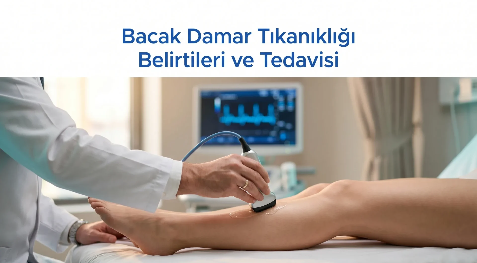
One of the complaints I encounter most frequently in my practice begins with this sentence: “Doctor, my leg seizes up when I walk, but it gets better when I sit down.” This is the classic early warning sign of leg vascular blockage. However, the disease does not always give such clear signals; sometimes it manifests as a sleepless night, sometimes as a small wound that refuses to heal, sometimes as nothing more than a cold foot.
In this article, I want to explain what we mean by “leg vascular blockage,” what symptoms it causes and what treatment options are available, in the same language I use with my patients. My aim is not only to provide information, but to help you find the right answer to the question “could this happen to me?”
What Is Leg Vascular Blockage?
Leg vessels fall into two main groups: arteries carry oxygenated blood from the heart to the legs, while veins return deoxygenated blood back to the heart. When people say “vascular blockage,” they usually mean the arterial system (Peripheral Artery Disease, PAD); however, Deep Vein Thrombosis (DVT) on the venous side is also a life-threatening form of blockage.
The underlying cause of arterial blockage is atherosclerosis, which involves the accumulation of plaques composed of fat and calcium on the inner wall of the vessel.1 As the plaque grows, the vessel lumen narrows, blood flow diminishes and the leg muscles do not receive sufficient oxygen. In venous blockage, the mechanism is different: a clot forms inside the vessel, disrupting venous return.
Quick note: “Leg vascular blockage” is commonly used in everyday language to refer to arterial disease; however, not every condition that causes leg swelling, colour change or pain originates from the arteries. Treating without a proper diagnosis is therefore incorrect.
The Difference Between Arterial and Venous Blockage
| Feature | Arterial (PAD) | Venous (DVT / Chronic Venous Insufficiency) |
|---|---|---|
| Pain character | Cramp-like pain that worsens with walking and subsides with rest | Burning and heaviness that worsen by end of day |
| Skin colour | Pale, bluish, cold | Reddish, brown pigmentation |
| Oedema | Rare | Frequent, prominent in the evenings |
| Pulse | Foot pulses weak or absent | Usually normal |
| Emergency risk | Critical limb ischaemia, amputation | Pulmonary embolism |
How Common Is Leg Vascular Blockage?
I receive this question frequently, and the answer usually surprises my patients. According to 2019 data, approximately 113 million people aged 40 and over worldwide are living with peripheral artery disease; global prevalence is 1.52%.2 According to NICE guidelines, the rate in the general population is 3-7%, rising to as high as 20% above the age of 60.3
In the National Treatment Guidelines of the Turkish Cardiovascular Surgery Society, the following figures are given for our country: asymptomatic PAD in the 50-60 age group is 2.5%; claudication (leg pain on walking) is 3% in those over 40 and 6% in those over 60.4 I can easily say from my own practice that these rates are increasing every year alongside an ageing population.
One more striking statistic: according to the CDC, 4 out of 10 patients with PAD experience no leg pain at all.5 In other words, the disease can be silent, which underlines the importance of regular check-ups.
Symptoms: Early, Intermediate and Advanced Stages
I tell my patients: “Our bodies start with small signals; if we don’t listen, they raise their voice.” Leg vascular blockage is no different. Grouping symptoms into three stages makes recognition easier.
Early Stage Symptoms
- Leg fatigue: Aching in the calf or thigh muscles over short distances
- Cold feet and legs: One-sidedness is particularly significant
- Thickening or brittleness of nails: An early sign of nutritional impairment
- Thinning or loss of leg hair
- Dry skin and pallor
Intermediate Stage: Intermittent Claudication
This term refers to cramp and pain in the leg after walking a certain distance, which subsides with rest. It is most commonly felt in the calf but can also affect the hip and thigh. My patients at this stage typically describe it in everyday terms such as “I have to stop every 100 metres on the way to the shops.”
Advanced Stage: Critical Limb Ischaemia
- Rest pain: Aching that appears when the leg is at rest, especially at night
- Non-healing wounds: Particularly around the toes and heel
- Purple, blue or black skin discolouration
- Signs of tissue loss (gangrene)
If any of these symptoms are present, there is no time to waste. Critical ischaemia requires intervention within 48-72 hours. Every hour of delay increases the risk of amputation.
Important: Leg pain that worsens at night and eases when the foot is dangled is the classic signal of severe arterial blockage. In this situation, you should consult the nearest vascular surgeon without delay.
Causes and Risk Factors of Leg Vascular Blockage
Risk factors for PAD largely overlap with those for cardiovascular disease. This also means that a patient with a blocked leg vessel is at significantly increased risk of heart attack and stroke.6
Modifiable Risk Factors
- Smoking: The strongest risk factor. Smokers have a 3-5 times higher risk of PAD
- Diabetes: Especially uncontrolled blood sugar, which damages the vessel wall
- Hypertension: Blood pressure causes micro-damage to the inner lining of vessels
- High cholesterol (LDL): The main component of plaque accumulation
- Obesity and a sedentary lifestyle
- Chronic kidney failure
Non-Modifiable Risk Factors
- Age: Risk rises rapidly after 50; prevalence above 70 is 14.5%
- Sex: Earlier onset in men; more pronounced in women after menopause
- Family history: History of early atherosclerosis in a first-degree relative
On the venous side, prolonged immobility (air travel, bed rest), hormone use, pregnancy, cancer and hereditary clotting disorders are the main factors. I covered this topic in detail in my article on DVT risk factors.
Diagnosis: Which Tests Do We Perform?
No single test is sufficient in the diagnostic process. In my clinic, the process works as follows:
1. Physical Examination
I carefully assess the leg pulses (femoral, popliteal, dorsalis pedis, posterior tibial). Skin temperature, colour, hair distribution, nail structure and capillary refill time all provide clues. In most patients, half the diagnosis is made here.
2. Ankle-Brachial Index (ABI)
This is the ratio of the systolic blood pressure at the ankle to that at the arm. Non-invasive, inexpensive and fast.7
| ABI Value | Interpretation |
|---|---|
| 1.0, 1.4 | Normal |
| 0.9, 1.0 | Borderline |
| 0.7, 0.9 | Mild PAD |
| 0.4, 0.7 | Moderate PAD |
| < 0.4 | Severe PAD, risk of critical ischaemia |
3. Doppler Ultrasound
Shows blood flow within the vessel, the site of stenosis and the flow velocity. Contains no radiation and can be repeated. It is indispensable for evaluating both arteries and veins.
4. CT Angiography and MR Angiography
Used when more advanced imaging is required and before surgical planning to map the vessels.
5. Conventional Angiography (DSA)
Referred to as the “gold standard.” A catheter is inserted at the groin and contrast medium is injected into the vessels. Balloon or stent placement can be performed in the same session (diagnosis and treatment combined).
Treatment Methods
Treatment is personalised for each patient. The stage of disease, lesion location, co-morbidities and the patient’s general condition are all determining factors. A stepwise approach can be summarised as follows:
1. Lifestyle Modifications
- Quitting smoking: The single most effective intervention. No medication replaces smoking cessation
- Supervised exercise programme: Supervised walking can increase claudication distance by an average of 180%
- Control of blood sugar, blood pressure and cholesterol
- Mediterranean-style diet
- Body weight control
2. Medical Treatment
- Antiplatelet agents: Aspirin or clopidogrel (reduce clot formation within vessels)
- Statins: Lower LDL cholesterol and stabilise plaques
- Cilostazol: For increasing claudication distance
- ACE inhibitors / ARBs: Hypertension control and vessel-protective effects
3. Endovascular Intervention (Minimally Invasive)
Today, the majority of PAD treatment consists of these techniques: balloon angioplasty, stenting, atherectomy, drug-eluting balloon. Access is gained through a small incision in the groin, and the patient is discharged the same day. Recovery time is very short.
4. Surgical Treatment
- Bypass surgery: A vascular graft is used to reroute blood flow around the blocked vessel
- Endarterectomy: Plaques are surgically removed from the inner lining of the vessel
- Thromboembolectomy: Urgently required in acute clot cases
On the venous side, my treatment approach differs: for varicose vein treatment, minimally invasive methods such as laser (EVLA), radiofrequency (RFA) and sclerotherapy are prioritised. For capillary vessel problems, you can find detailed information on our capillary vein treatment page.
When Should You Go to the Emergency Department?
If the following symptoms are present, time is critical:
- Sudden severe pain and colour change in the leg (whitening, purpling)
- Sudden loss of sensation or muscle weakness in the leg
- Inability to feel a pulse on one side
- A rapidly enlarging wound or black patch
- Sudden swelling in one leg accompanied by shortness of breath (suspected pulmonary embolism)
These presentations may indicate acute arterial occlusion or DVT-related pulmonary embolism. Both are life-threatening. Go to the emergency department without delay.
What Happens If Left Untreated?
The answer varies by disease, but the common ground is this: vascular blockage is not a self-resolving condition. Untreated PAD can progress to critical limb ischaemia, non-healing wounds and ultimately amputation. Furthermore, the 5-year mortality rate for PAD patients is 15-30%; when I see this figure I tell my patients: “Vascular health is an indicator of life expectancy.”8
Frequently Asked Questions
Can leg vascular blockage resolve?
Plaques are not fully reversible, but their progression can be halted and symptoms largely eliminated. I very frequently see patients who have quit smoking, walk regularly and adhere to drug treatment show a halt in disease progression.
What does leg vascular blockage pain feel like?
Typically cramp-like pain that starts during walking and subsides within 5-10 minutes of rest. In advanced stages it can occur at night during rest and eases when the foot is dangled.
Which doctor treats leg vascular blockage?
A Cardiovascular Surgery specialist evaluates both the arterial and venous sides and manages diagnosis and treatment.
What do I feel during an ABI test?
Nothing. Blood pressure cuffs are placed on the ankle and arm, and the pulse is detected with a Doppler probe. It is a 15-minute, painless procedure. The diagnostic information gained is extremely valuable.
Does walking exercise really work for vascular blockage?
Absolutely. Studies show that supervised walking programmes can be as effective as balloon angioplasty in some patients. Walking 3 days a week, 30-45 minutes at a time, until pain is felt and then resting for 2-3 minutes, makes a significant difference after 3 months.
Are varicose veins and vascular blockage the same thing?
No. Varicose veins are a problem of the venous system, while blockage is most often a problem of the arterial system. I clarified this distinction in my article on what are varicose veins and why do they occur.
Is leg vascular blockage related to nocturnal leg pain?
Rest pain at night is typical in advanced PAD. But nocturnal leg pain has many other causes (restless leg syndrome, cramps, neuropathy). I covered the various sources in detail in my article on causes of night leg pain.
Can blockage recur after treatment?
Yes, especially if underlying risk factors are not brought under control. In a patient who continues to smoke, even a stent can become blocked within two years. Treatment is not just an intervention, it is a complete process combined with lifestyle change.
Final Words
When caught early, leg vascular blockage can usually be controlled with minimally invasive methods. The real danger is postponing for years by attributing the pain to “old age.” If you are experiencing several of the symptoms I have described above, do not delay seeing a vascular surgeon. Rather than worrying without a diagnosis, you can get a clear answer with an objective ABI and Doppler evaluation.
Op. Dr. Onur Üstünel
Cardiovascular Surgery Specialist
For international patients: If you are considering chronic venous insufficiency treatment in Turkey, our İzmir clinic provides comprehensive evaluation, Doppler ultrasonography and personalized treatment planning. Review our treatment overview for protocols and pricing. We also offer expert management of peripheral artery disease and deep vein thrombosis.
References
- CDC. About Peripheral Arterial Disease (PAD). https://www.cdc.gov/heart-disease/about/peripheral-arterial-disease.html
- Song P, et al. Global burden of peripheral artery disease and its risk factors, 1990-2019. PMC10522777. https://pmc.ncbi.nlm.nih.gov/articles/PMC10522777/
- NICE Clinical Guideline CG147. Peripheral arterial disease: diagnosis and management. https://www.nice.org.uk/guidance/cg147
- Türk Kalp ve Damar Cerrahisi Derneği. Periferik Arter ve Ven Hastalıkları Ulusal Tedavi Kılavuzu 2021. https://www.tkdcd.org/content/887/periferik-arter-ve-ven-hastaliklari-ulusal-tedavi-kilavuzu-2021
- NHLBI (NIH). Peripheral Artery Disease. https://www.nhlbi.nih.gov/health/peripheral-artery-disease
- American Heart Association. What Is Peripheral Artery Disease? https://www.heart.org/en/health-topics/peripheral-artery-disease/about-peripheral-artery-disease-pad
- Epidemiology of Peripheral Artery Disease: Narrative Review. PMC9320565. https://pmc.ncbi.nlm.nih.gov/articles/PMC9320565/
- Peripheral Arterial Disease. StatPearls, NCBI Bookshelf. https://www.ncbi.nlm.nih.gov/books/NBK430745/